

Portable Ultrasound Machine KUS-A200 is a compact B/W digital ultrasound imaging modality equipped with 10” LCD screen and 6 imaging modes. Core features include digital beam-forming, ultrasonic frame-forming, real-time aperture control, dynamic focusing and gamma correction to ensure efficient measuring and spot-analysis of the body area under study.
| Display Size | 10-inch screen |
| Display Depth | ≥ 250 mm |
| Ultrasound Modes | B, B+B, B+M, B+2M, M, 4B |
| TGC | Total gain, 8 segment TGC |
| Depth Adjustability | 16 level of real-time depth adjustments |
| Probe Scan Methods | Convex, Linear, Micro-convex |
| Cine-loop | ≥ 400 frames |
| Frame Storage | ≥ 64 frames |
| Body Marks | 40 types |
| Imaging Mode | 2D ultrasound imaging |
| Measurement | Distance, Circumference, Area and Volume |
| Cardiac Measurement | LV/VF, AV, MV, PV, TV, HR |
| Obstetric Measurement | BDP, FL, AC, HC, CRL, AD, GS, G.A, LMP, EDD, FW |
| Image Processing | Pseudo-color, Dynamic range, THI, Image Smoothing, Image Sharpening, Gamma Correction and Histogram based Enhancement. |
| Zoom | 4 step zoom-in functionality |
| Output Interface | PAL-D Video, RS-232, USB 2.0 |
| Dimensions | 460 x 360 x 430 mm |
| Weight | 9 Kgs |
Used in examination rooms, consultation clinics, primary healthcare centers and rescue ambulance or outdoor examinations for point of care ultrasonography imaging for symptomatic examination and diagnosis at patient’s bedside.
| Sr. | Standard Accessories | Qty |
|---|---|---|
| 1 | 3.5 MHz Convex Probe | 1 |
| Sr. | Optional Accessories | Qty |
|---|---|---|
| 1 | 7.5 MHz Linear Probe | 1 |
| 2 | 5.0 MHz Micro-convex Probe | 1 |
| 3 | 6.5 MHz Transvaginal Probe | 1 |
| 4 | 7.5 MHz Endorectal Probe | 1 |
Used in examination rooms, consultation clinics, primary healthcare centers and rescue ambulance or outdoor examinations for point of care ultrasonography imaging for symptomatic examination and diagnosis at patient’s bedside.

Display Size : 12-inch LCD screen
Display Depth : 40mm to 250 mm
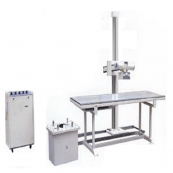
Exposure Voltage50 kV ~ 90 kV'
Exposure CurrentSmall focus: 25 mA </br> Big focus: (50, 75, 100 and 125) mA'
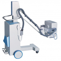
Inverter Frequency50 kHz'
Tube Voltage40 kV – 100 kV (interval 1 kV)'
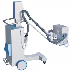
Inverter Frequency50 kHz'
Tube Voltage40 kV – 120 kV (interval 1 kV)'

Inverter Frequency50 kHz'
Tube Voltage40 kV – 120 kV (interval 1 kV)'

Max Output Power4 kW'
Nominal Power100 kV, 40mA, 0.1 s, 4 kW'

Focal Distance430 × 430 mm'
Source Head Range550 mm - 1820 mm'
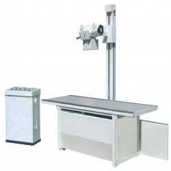
Exposure Voltage50 kV ~ 125 kV'
Exposure CurrentSmall focus: (50, 100) mA </br> Big focus: (50, 100, 200 and 300) mA'

Focal Distance 350 x 350 mm'
Source Head Range520 mm - 1880 mm'

Exposure Voltage50 kV ~ 100 kV'
Exposure CurrentSmall focus: 50 mA </br> Big focus: (50, 100, 150 and 200) mA'

Generator TypeHigh frequency inverter 80 kHz'
Radiographic RatingsLarge focal point, 20-35 kV/10-510 mAs <br/> Small focal point, 20-35 kV/ 10-110 mAs'

Generator TypeHigh frequency inverter 40 kHz'
Radiographic RatingsLarge focal point, 20-35 kV/10-510 mAs <br/> Small focal point, 20-35 kV/ 10-110 mAs'

No. of Channels10'
No. of Sample Analysis5'

Tube Voltage60 kV'
Tube Current8 mA'

Tube Voltage90 kVp'
Tube Current10 mA'

Tube Voltage60 kV'
Tube Current1 mA'

Tube Voltage60 kV'
Tube Current8 mA'

Image Sensor1/4″ Super HAD color CCD'
CCD Resolution (HD optional)8,00,000 pixels'

Display3” TFT panel, 4:3 ratio'
MagnificationOptical : 20-200 X </br> Digital : 500 X'

LCD Display6” Real-time display'
Expandable StorageUp to 1008 ECGs'

LCD Display6” LCD real-time'
Expandable StorageUp to 250 ECGs'

LCD Display192 x 64 Graphic'
Expandable Storage'

LCD Display3.8 inch Graphic'
Expandable StorageUp to 250 ECGs'

Number of channels4 channel'
Stimulus ModesSingle-current, Dual-current Stimulator'

BulbOne spare bulb'
Bulb typeStainless bulb'

Otoscope1'
Size ear speculum3'

Light sourceLED bulb'
LuminanceAbout 18.5 lumen'

TypeTwo-channel for independent outputs'
Test SignalPure tone, Pulse tone, Warble tone, Narrowband noise'

Flow range1 L/s ⁓ 16 L/s'
Volume range10 L'

Input channel(12/24/36/48 Monopolar EEG+12channelsBipolar EEG+12channels PSG)'
Recording modesReal-time'

Input channel(19 Monopolar EEG and 1 Bipolar ECG)'
Recording modesReal-time (via Bluetooth) / Dynamic (via Flash memory)'

Input channel32/64/128 channels Monopolar EEG'
Recording modesReal-time'

Range of measurement<table class="tableizer-table"> <thead><tr class="tableizer-firstrow"><th>Lens</th><th>Range</th></tr></thead><tbody> <tr><td>Sphere</td><td>-25 ~ +25 D unit( 0.01/0.12/0.25 D steps)</td></tr> <tr><td>Cylinder</td><td>-9.99 ~ +9.99 D unit (0.01/0.12/0.25 D steps)</td></tr> <tr><td>Axis</td><td>0° ~ 180° unit (1° steps)</td></tr> <tr><td>Add</td><td>0 ~ +9.99 D unit (0.01/0.12/0.25 D)</td></tr> <tr><td>Prism</td><td>0 ~ 6 △ unit ( 0.01/0.12/0.25 △)</td></tr> </tbody></table>'
Lens measurement type<table class="tableizer-table"> <thead><tr class="tableizer-firstrow"><th>Lens</th><th>Type</th></tr></thead><tbody> <tr><td>Cylinder</td><td> +,+/-,-</td></tr> <tr><td>Prism</td><td> X-Y,P-B</td></tr> <tr><td>Contact lens </td><td> Soft/hard</td></tr> </tbody></table>'


